

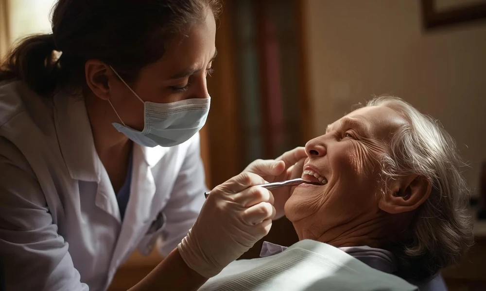
You go in for a routine dental visit with a dentist in Montclair, NJ, expecting to hear about plaque, a filling, or maybe gum care, and instead your dentist points out a white patch or a red area in your mouth. That can stop you cold. Most people have never heard the words erythroplakia or leukoplakia until they are sitting in the chair, trying to read their dentist’s face and wondering if this means cancer.
That fear is real, and the uncertainty is often the hardest part. These terms describe oral tissue changes that need attention, not panic. Some patches turn out to be harmless irritation. Some need monitoring. Some need a biopsy because certain red or white lesions in the mouth can carry a higher risk of precancer or cancer. The point is not to ignore them and not to assume the worst. A general and family dentist may be the first person to spot these changes, which gives you a chance to act early.
Leukoplakia and erythroplakia describe visible oral tissue changes
Leukoplakia usually refers to a white patch in the mouth that cannot be rubbed off and cannot be explained by another clear cause right away. Erythroplakia refers to a red patch that also does not have an obvious simple explanation. Red and white mixed areas can appear too, and those may also need close evaluation.
The reason dentists pay attention to these lesions is simple. Color changes in the lining of the mouth can reflect irritation, infection, inflammation, or abnormal cell changes. Erythroplakia often raises more concern than leukoplakia because red lesions are more likely to show dysplasia or cancerous change when biopsied. That does not mean every red patch is dangerous. It means it deserves a careful look.
You might be thinking back to a spot you noticed weeks ago, maybe one that did not hurt, which made it easy to dismiss. That happens all the time. Many oral tissue changes cause little or no pain at first. A sore area that lingers, a thickened patch, a rough spot, or a color change that does not fade should not be brushed off just because it is not dramatic.
Some mouth lesions come from irritation, but persistent patches need follow up
Denture friction, a rough tooth edge, tobacco use, alcohol use, chronic cheek biting, and some infections can all change the look of oral tissues. If there is an obvious irritant, your dentist may address that first and recheck the area soon after. If the patch remains, the next step may be referral for further testing or a biopsy.
This is where people often get stuck. They want a fast yes or no answer, but oral lesions do not always work that way. A dentist may say, “We need to watch this for two weeks,” and that waiting period can feel long. It is not a brush off. It is often a way to see whether the tissue returns to normal once the likely cause is removed.
If a lesion persists, looks suspicious, or has features linked with higher risk, your dentist may recommend an oral cancer screening approach and possible biopsy. The National Cancer Institute explains how oral cancer screening and follow up are used to evaluate abnormal findings. If cancer is diagnosed, treatment depends on the size, location, and stage of the lesion. The NCI also outlines lip and mouth cancer treatment options.
Watching and testing serve different purposes
People sometimes hear “watch it” and assume that means “ignore it.” It does not. Monitoring is an active plan with a timeline. Biopsy is a diagnostic step used when the appearance, location, or persistence of the lesion calls for tissue testing. Both approaches have a place.
| Approach | When it may be used | What it helps with | Limits |
|---|---|---|---|
| Short term monitoring | When there is a likely irritant such as denture rubbing or cheek biting | Shows whether the tissue heals after the cause is removed | Does not confirm what the cells look like under a microscope |
| Biopsy | When a patch persists, looks suspicious, or has high risk features | Can identify dysplasia, precancer, or cancer | Requires a procedure and pathology review |
| Routine dental exams | For ongoing surveillance, especially if you have risk factors | Helps catch new or changing lesions early | Cannot replace biopsy when a lesion needs diagnosis |
The difference matters because a white patch from friction may improve once the source is corrected, while a persistent red lesion may need tissue sampling even if it does not hurt. That is why terms like white patches in the mouth and red lesions are descriptive, not final diagnoses.
Practical steps help you respond without spiraling
1. Track what you see.
Take a clear photo if the spot is visible, note when you first noticed it, and write down any symptoms such as burning, bleeding, tenderness, or change in size. If you use tobacco, vape, drink heavily, wear dentures, or recently had dental irritation, include that too. These details help your dentist compare changes over time.
2. Remove obvious irritants and keep the recheck appointment.
If your dentist adjusts a denture, smooths a rough edge, or asks you to stop using a product that may be irritating your mouth, follow through. Then return when asked. A lesion that stays after the irritant is gone tells a different story than one that heals quickly.
3. Ask direct questions about next steps.
Ask whether the area looks low risk or suspicious, how long it should take to improve, and what would trigger a biopsy or referral. If you are told to monitor it, ask for a specific timeline. If a biopsy is recommended, ask what type is planned and when results usually come back. Clear answers lower the stress because you know what happens next.
Early attention to oral mucosal lesions gives you more control
Hearing unfamiliar terms at the dentist can make your mind race, but the most useful response is steady follow up. Erythroplakia and leukoplakia are signs your dentist is paying close attention to changes that many people would miss on their own. That is a good thing. The goal is to sort out irritation from something more serious before time is lost.
If your dentist mentions an abnormal oral lesion, take it seriously, keep the follow up plan, and ask for clear guidance on whether monitoring or biopsy makes sense. A general and family dentist can play a key role in spotting these changes early and helping you move to the next step with less fear and more clarity.
